May 3 2017
 This is a schematic of an achromatic X-ray microscope based on total-reflection mirrors. Credit: Osaka University
X-ray microscopes are mostly used together with full-field imaging techniques in spectromicroscopy applications, where they permit the chemical structures of materials to be examined and visualized simultaneously.
The performance of these microscopes is frequently affected by issues with chromatic aberrations, referring to optical effects that bring about limitations to the resolution or degree of fineness to which images of the material structures can be obtained. Earlier solutions to the problem have frequently proved to be complicated to manufacture and implement. A collaborative team headed by researchers from Osaka University has thus created an optical system capable of being used in full-field X-ray microscopes that provides a more practical solution to overcome the chromatic aberration problem.
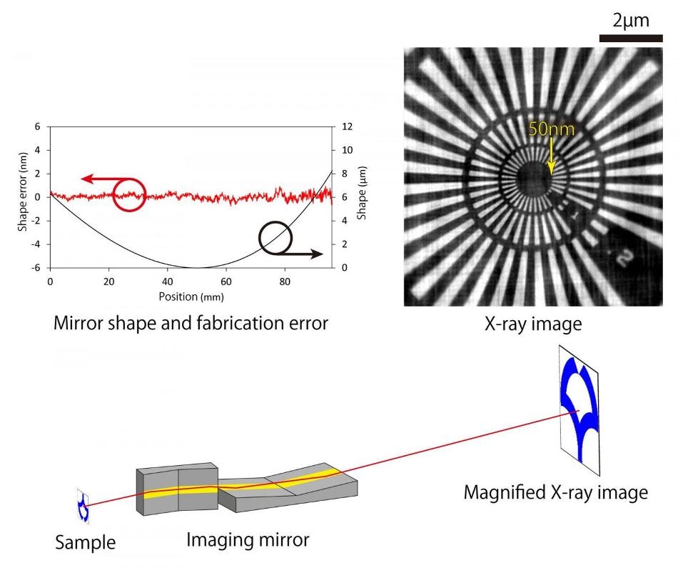
We developed an imaging optical system based on use of two monolithic imaging mirrors. These mirrors have elliptical and hyperbolic shapes on a single substrate, and fixing of the relative positioning between the ellipse and the hyperbola can provide high image quality with lasting stability.
Satoshi Matsuyama, Assistant Professor, Graduate School of Engineering, Osaka University
Fabrication of this complex mirror system pointed out the necessity to alter the existing manufacturing processes, but the proposed mirror structures were developed with the desired shapes to an accuracy of almost 1 nm.
A custom designed alignment system was used to assemble the mirror structure, and after this step it was employed in a full-field X-ray microscope system for performance testing at the Spring-8 synchrotron radiation facility.
The microscope was tested for its spatial resolution, the presence of chromatic aberrations, and long-term stability using a fine test pattern called a Siemens star and a photon energy of approximately 10 keV. We were able to clearly resolve 50-nm-sized features with high stability over a period of 20 hours without any chromatic aberrations.
Professor Kazuto Yamauchi, Center for Ultra-Precision Science and Technology, Osaka University
The developed system was implemented in X-ray absorption fine structure spectromicroscopy experiments, and the system succeeded in identifying both chemical states and elements in micron-sized specimens of tungsten and zinc. This system will be used in future research in order to enhance its performance towards the theoretical limit, and it already proves to be promising for use in a variety of applications, including high-resolution full-field X-ray fluorescence imaging and ultra-fast imaging with high-intensity X-rays. It could also be possible to use this mirror structure in various other systems, with promising applications such as focusing and imaging optics for synchrotron radiation X-rays and X-ray-free electron lasers.