Aug 22 2016
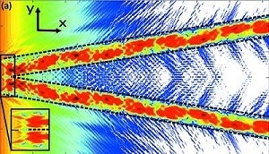 Dashed lines mark the channel boundaries and the enlarged part displays the region where the wave is coupled in. The intensity is normalized and plotted in logarithmic scaling. (Credit: Hoffmann-Urlaub and Salditt)
Filtering, coupling, guiding, confining, or splitting beams of visible light is often performed using waveguides. However, developing waveguides that are capable of doing the same for X-rays is quite challenging in fabricatio, which is the reason they are still in the early stages of development.
In the current issue of Acta Crystallographica Section A: Foundations and Advances, Sarah Hoffmann-Urlaub and Tim Salditt illustrate the fabrication and testing of a millimetre-sized chip capable of splitting a beam of X-rays.
Fork-shaped channels measuring just a few tens of nanometers in width and depth are moved into a silicon wafer with the help of electron-beam lithography and reactive ion etching, then enfolded by bonding another silicon wafer on top.
The outcomes of simulations of how the 'parent' beam is divided into two 'daughter' beams on passing via the chip were verified by experimental measurements at the European Synchrotron Radiation Facility, revealing that the incident beam is properly transported through the chip, carefully divided and guided to exits that have precisely controlled and tunable spacings.
Once the daughter beams leave the chip, they interfere, causing a pattern of vertical stripes similar to the pattern procured from a classical Young's double-slit interference experiment. On close examination, fork-like structures existed within the stripes that start off from discontinuities in the phase of the recombined beam, generating striking characteristics referred to as phase vortices.
From those interference patterns the intensity dispersion in the exit plane of the channels is rebuilt, which is discovered to be in excellent agreement to the real channel design.
This research complements a previous study on 2D confined channels in silicon in straight and tapered geometries, and facilitates the way to accomplishing `X-ray optics on a chip'.
When the samples are illuminated by the two beams, it could lead to some appealing benefits for coherent imaging and makes way for a potentially new form of nano-interferometer.
Going forward the researchers aim to further develop their beamsplitter to generate many daughter beams from a single parent beam, which would enable an object to be imaged concurrently by a number of beams from various directions.
Source: http://www.iucr.org