Nov 18 2015
A plethora of images from live-bearing brittle stars allows a 3D look inside these unique starfish relatives and an insider's view of their soon-to-be offspring
 GigaScience 2015: 4. doi:10.1186/s13742-015-0093-2.
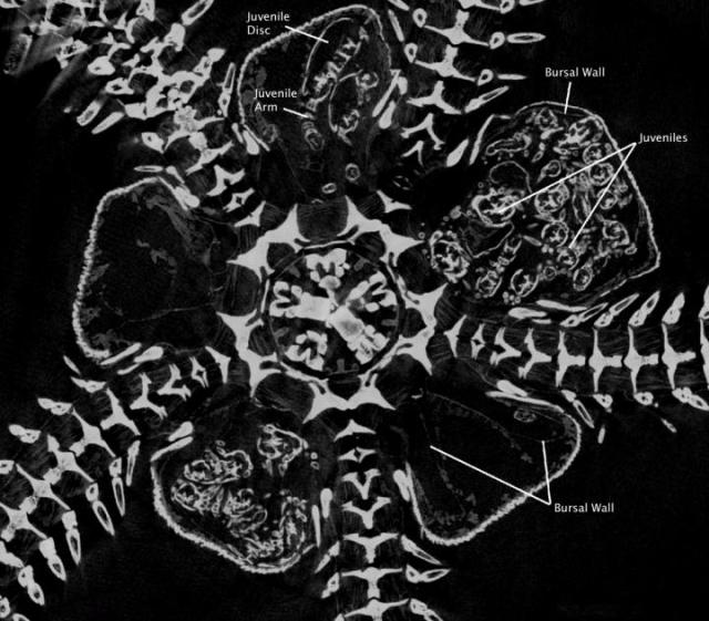
Published today in GigaScience is an article that describes high-resolution 3D images, data, and videos of five individuals from three different species of live-bearing brittle stars [1]. The entire associated 100GB of data is freely available in the GigaScience database, GigaDB [2]. These data, generated using non-invasive micro X-ray computed tomography (microCT), were used to study the development of young brooded inside the brittle star [3], but the value of these data -- besides being cool to look at -- is in their suitability for broad sharing for other researchers to use to examine juveniles within the adult as well as to carry out comparative morphological or anatomical analyses. Large-data sharing is an essential part of making research both reproducible and reusable in the broadest sense. Enabling better understanding of the complex interplay between internal structures can also help throw new insight into how species interact with their environments.
Brittle stars are a class of organisms that are closely related to starfish. While most members of this group reproduce externally, there are some species that develop their young internally, and then 'give birth' to live young. While sonograms are the means by which humans can get a look at their developing offspring, live-bearing brittle stars have not been so lucky, as researchers primary means to investigate the brooding chambers and juveniles of brittle stars has been via dissection. This technique destroys the sample, thus eliminating the possibility of further study and sharing of samples for others to use. Here, by using microCT scanning, the authors were able to visualize in 3D the brooding chambers (bursae) and juveniles inside the brittle star, providing an in situ view of the developing young and the position of the bursae inside the adult. Further, because the process doesn't damage the brittle star, additional analyses can be carried out on the same individual.
First author Jannes Landschoff from the Applied Marine Science Institute at the University of Cape Town states: "Our goal was to visualise the very large brooded juveniles inside the adults without disturbing the surrounding tissues. At first, we purely wanted to show the feasibility, but we soon realised how amazing the three-dimensional visualisations looked and that our rotation movies attracted much interest". Recognizing the extremely high quality of the images they used to study brittle star development, and that the data included much more information than they actually used, they made all these data available to others in a fully open-access format through GigaScience's repository, GigaDB. The data includes videos and projection, reconstruction images and an additional volume file for easy viewing.
Source: http://www.gigasciencejournal.com/