Nov 15 2012
Doctors can detect evidence of heart disease faster than ever before using new 3-D nuclear imaging technology at the University of Rochester Medical Center’s Heart and Vascular Center.
 The new D-SPECT nuclear cardiology camera is a major advance in providing accurate, high-speed imaging of heart function.
The high-speed D-SPECT digital gamma camera system provides “amazing images with a fraction of the radiation dose,” said Ronald G. Schwartz, M.D., M.S., director of Nuclear Cardiology. “This camera is a huge leap forward from all other available technology for accuracy, speed, comfort and safety for our patients and providing ultra, low-dose imaging capability.”
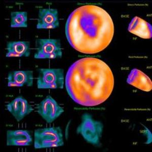
A single-photon emission computerized tomography (SPECT) camera is used to measure heart muscle blood flow and function for patients with heart disease or suspected heart problems. This very popular technique uses a safe, very low dose of radioactive tracer to create 3-D images or “moving maps,” that show the blood flow through the heart muscle and heart chambers.
.“These moving maps can be rotated to carefully examine every square inch of the heart muscle to look for damage or jeopardized areas,” said Schwartz, the region’s most experienced nuclear cardiologist and past president of the Cardiovascular Council of the Society of Nuclear Medicine.
Patients can now be evaluated very safely with a fraction of the radiation dose available by other invasive and non-invasive x-ray coronary imaging methods and without the use of contrast agents, a known risk for patients with kidney disease.
For patients who cannot exercise and require a medication stress test, D-SPECT continues to offer important safety advantages. “The stress medication, regadenoson, is the most widely prescribed pharmacologic stress agent, and has been proven safe for patients with advanced stages of kidney disease, asthma and emphysema in recent large clinical trials,” Schwartz said.
The D-SPECT system, located in the Paul Yu Heart Center, offers a 10-fold increase in sensitivity and enhanced spatial resolution, similar to cardiac PET scan. The enhanced sensitivity allows nuclear cardiologists to reduce the amount of radioactive material to ultra-low dose levels never before achieved. Obese patients, as heavy as 540 pounds, can be imaged with high clarity and accuracy of diagnostic information, which has never been possible with this or other cardiac imaging techniques.
Results of personalized, patient-centered imaging using D-SPECT technology allows cardiologists to determine the need for heart catheterization, angioplasty or bypass surgery, or medications for patients with heart disease. In addition, nuclear imaging helps doctors evaluate the effectiveness of these therapies.
“We have already begun to take advantage of the remarkable efficiency of the D-SPECT nuclear cardiology camera to assess patients suspected of having heart disease in under an hour,” said Schwartz, professor of Medicine and Imaging Sciences.
“Our patients and doctors are very happy with this added capability. The D-SPECT camera makes the experience much better during a stressful time,” said Maria Mackin, M.S., C.N.M.T., R.T.(N), chief technologist and supervisor of Nuclear Cardiology laboratories.
The open design of the scanner allows patients to sit in a reclining chair, similar to those in a dentist’s office, and the camera is positioned in front of the patient’s chest to capture the images. It is a significant benefit for people who experience claustrophobia during similar imaging tests that involve passing through a donut-shaped scanner.
The D-SPECT system is only available at a handful of other leading institutions across the country, including the Mayo Clinic and Harvard, Yale, Southwestern, Mid-American and Cedars Sinai medical centers.
Schwartz expects the technology will support URMC’s heart research into the benefits of specialized pacemakers in heart function, measurement of absolute coronary blood flow and improving ways to prevent heart failure after chemotherapy for cancer.
Source: http://www.urmc.rochester.edu