Posted in | News | Imaging | Medical Optics

Acquire 3D Nerve Cell Images of the Brain without the Need for Sectioning

The completely redesigned plan-apochromatic 20x/1.0 VIS-IR objective lens from Carl Zeiss now makes it possible to acquire 3D images of nerve cells down to depths of 5.6 mm in intact tissue using a confocal laser scanning or multiphoton microscope, without having to section the brain.
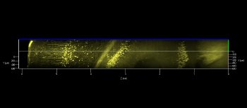
This permits the three-dimensional visualization of the branches of individual nerve cells and the imaging of their connections. In untreated tissue, the penetration depth now achievable is five to ten times more that of a multiphoton microscope featuring traditional optics.

Using the microscope to visualize and track nerve cells deep into the brain is an important step in the decoding of the brain circuitry. The new objective lens from Carl Zeiss now brings this goal considerably closer to realization. Carl Zeiss has designed the plan-apochromatic 20x/1.0 VIS-IR objective lens for the clearing method known as Scale developed by Dr. Atsushi Miyawaki at the RIKEN Brain Science Institute in Japan. A special water-based reagent solution transforms the sheath substance of the nerve cells into a transparent matrix without impairing the signals from fluorescent marker and tracer substances.

“I am very impressed by the new possibilities,” says Dr. Atsushi Miyawaki, whose results have been published in Nature Neuroscience and provoked broad interest in the neurosciences. In one of the most important research projects in the field of brain research, the Connectome project, the objective lens will help scientists obtain a better understanding of brain connectivity.

A prototype of the objective lens was presented at Neuroscience 2011 in Washington D.C. After concluding system tests, it will then become available for ZEISS confocal and multiphoton microscopes in 2012.

Citations

Please use one of the following formats to cite this article in your essay, paper or report:

  • APA

    Carl Zeiss Microscopy GmbH. (2019, February 28). Acquire 3D Nerve Cell Images of the Brain without the Need for Sectioning. AZoOptics. Retrieved on April 18, 2024 from https://www.azooptics.com/News.aspx?newsID=15367.

  • MLA

    Carl Zeiss Microscopy GmbH. "Acquire 3D Nerve Cell Images of the Brain without the Need for Sectioning". AZoOptics. 18 April 2024. <https://www.azooptics.com/News.aspx?newsID=15367>.

  • Chicago

    Carl Zeiss Microscopy GmbH. "Acquire 3D Nerve Cell Images of the Brain without the Need for Sectioning". AZoOptics. https://www.azooptics.com/News.aspx?newsID=15367. (accessed April 18, 2024).

  • Harvard

    Carl Zeiss Microscopy GmbH. 2019. Acquire 3D Nerve Cell Images of the Brain without the Need for Sectioning. AZoOptics, viewed 18 April 2024, https://www.azooptics.com/News.aspx?newsID=15367.

Tell Us What You Think

Do you have a review, update or anything you would like to add to this news story?

Leave your feedback
Your comment type
Submit

While we only use edited and approved content for Azthena answers, it may on occasions provide incorrect responses. Please confirm any data provided with the related suppliers or authors. We do not provide medical advice, if you search for medical information you must always consult a medical professional before acting on any information provided.

Your questions, but not your email details will be shared with OpenAI and retained for 30 days in accordance with their privacy principles.

Please do not ask questions that use sensitive or confidential information.

Read the full Terms & Conditions.